
One of the fundamental tools of neuroscience is quantifying the amount of various molecules in the different regions of the brain. These molecules can be protein, radioactive tracers, DNA/RNA, or others depending on the imaging modality. Regardless of the modality used, the region(s) in which the molecule should be quantified needs to be defined. These regions of interest (ROI) can be created by manual delineation using software such as ITK-SNAP or ImageJ. However, this process is time-consuming and prone to inter- and intra-operator variability. So, methods to automatically create these regions (a process called segmentation) are of great interest.
|
DAPI template Running back and forth |
 |
For magnetic resonance imaging (MRI) and computed tomography (CT), a method called template-based segmentation is widely used. First, a population-based template is created by taking images of several individuals, aligning them (a process called image registration), and then taking the average of all the images. The average image is called the template and is segmented by hand multiple times by different experts. A new brain image acquired with the same imaging technique can then be aligned/registered to the template and thus segmented automatically.
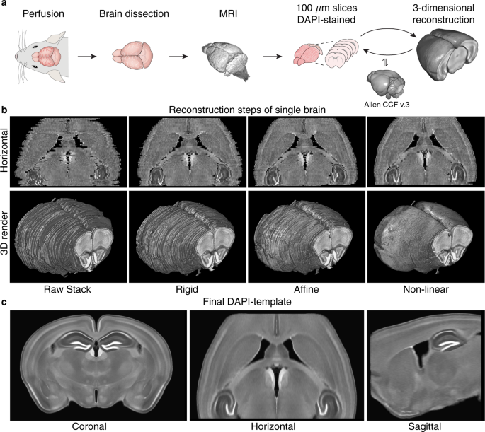
In this study, we sought to allow for template-based segmentation of 2-dimensional mouse brain slices acquired with fluorescence microscopy. Typically, a stain called DAPI is used to show the brain structures, and up to three additional channels are used for measuring the molecule(s) of interest. So, we needed to construct a population-based template of the mouse brain from slices stained with DAPI.
We collected consecutive coronal slices from 12 mice (C57BL/6) covering the majority of the cerebrum and DAPI-stained them. With the help of Allen Mouse Brain Common Coordinate Framework (CCFv3) we were able to reconstruct all 12 brains. This was done by iteratively stacking the 2-dimensional slices into 3-dimensional volumes, registering, and slicing. With the reconstructed brains, we could perform the template creation in 3-dimensional space. Using this new template in combination with the CCFv3 we started over with the reconstruction and template creation resulting in the final well-defined DAPI template (shown in the beginning of the post).

We were thrilled with how well the reconstruction process worked and happy to see a well-defined and smooth template. New coronal slices were easily registered to the template, so we wrote a small python program for automatic template-based segmentation (see the full paper for examples). To encourage the scientific community to utilize and improve the template, we have made all the data available in a well-structured and ordered fashion here: https://doi.org/10.12751/g-node.16wrxa
The full paper including a thorough explanation of the reconstruction and validation process can be found here: nature.com/articles/s41597-020-0570-z


Please sign in or register for FREE
If you are a registered user on Research Communities by Springer Nature, please sign in